An international study led by Ohio State University neuroscience researchers describes one of the missing triggers that controls calcium inside cells, a process important for muscle contraction, nerve-cell transmission, insulin release and other essential functions. The research is being posted online April 22 in the journal Nature. The researchers believe the findings will enhance the understanding of how calcium signals are regulated in cells and shed light on new ways to treat many diseases, including cardiovascular diseases, immune diseases, metabolic diseases, cancer, and brain disorders.
The study found that molecular structures called two-pore channels (TPCs) cause the release of calcium when stimulated by a substance called NAADP. The researchers also show that TPCs are located in the membranes of cell components called lysosomes and endosomes. These are mobile structures within cells that were not previously thought to be sites of calcium release. Furthermore, the discharge of calcium from these structures can prompt much larger releases from stores located on the large and elaborate membrane network called the endoplasmic reticulum.
“Our study discovered one of the missing targets for calcium signaling,” says Michael Xi Zhu, associate professor of neuroscience and a researcher with Ohio State’s Center for Molecular Neurobiology. “It also nails down that NAADP receptors are located on lysosomes and endosomes, which should change people’s views of calcium signaling. “It’s as if we now understand that cells have not only a primary battery for calcium but other batteries in different places.” Researchers have known for some time that NAADP, or nicotinic acid adenine dinucleotide phosphate, stimulates calcium release inside cells, but there was controversy about how this happened and where this calcium source was located.
Zhu, working with colleagues at the University of Edinburgh, the University of Oxford and UMDNJ-Robert Wood Johnson Medical School in New Jersey, used gene sequence information to discover first that TPC proteins should have the properties of a calcium channel. The investigators tested their hypothesis in a series of experiments that involved boosting TPC levels – specifically, TPC2 – in a line of laboratory cells. They found that higher TPC2 levels corresponded to higher calcium levels in cells exposed to NAADP.
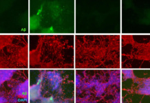
They used fluorescent antibody labeling to show that the TPC proteins are localized in the membranes of lysosomes and endosomes, which are two types of vesicles in cells. Lysosomes contain enzymes that digest materials and kill bacteria, while endosomes contain materials taken up from the external environment and internalized. Finally, the researchers found that these NAADP-sensitive stores of calcium are tightly coupled to the larger calcium stores on the endoplasmic reticulum.
This work was supported by grants from the U.K. Wellcome Trust, British Heart Foundation, U.S. National Institutes of Health and American Heart Association. Adapted from materials provided by Ohio State University Medical Center, via EurekAlert!, a service of AAAS.


