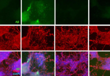
In the photo to left, we see young, healthy muscle (left column) appears pink and red. In contrast, the old muscle is marked by scarring and inflammation, as evidenced by the yellow and blue areas. This difference between old and young tissue occurs both in the muscle’s normal state and after two weeks of immobilization in a cast. Exercise after cast removal did not significantly improve old muscle regeneration; scarring and inflammation persisted, or worsened in many cases.
A study led by researchers at the University of California, Berkeley, has identified critical biochemical pathways linked to the aging of human muscle. By manipulating these pathways, the researchers were able to turn back the clock on old human muscle, restoring its ability to repair and rebuild itself. The findings will be reported in the Sept. 30 issue of the journal EMBO Molecular Medicine, a peer-reviewed, scientific publication of the European Molecular Biology Organization.
“Our study shows that the ability of old human muscle to be maintained and repaired by muscle stem cells can be restored to youthful vigor given the right mix of biochemical signals,” said Professor Irina Conboy, a faculty member in the graduate bioengineering program that is run jointly by UC Berkeley and UC San Francisco, and head of the research team conducting the study. “This provides promising new targets for forestalling the debilitating muscle atrophy that accompanies aging, and perhaps other tissue degenerative disorders as well.”
Previous research in animal models led by Conboy, who is also an investigator at the Berkeley Stem Cell Center and at the California Institute for Quantitative Biosciences (QB3), revealed that the ability of adult stem cells to do their job of repairing and replacing damaged tissue is governed by the molecular signals they get from surrounding muscle tissue, and that those signals change with age in ways that preclude productive tissue repair.
Those studies have also shown that the regenerative function in old stem cells can be revived given the appropriate biochemical signals. What was not clear until this new study was whether similar rules applied for humans. Unlike humans, laboratory animals are bred to have identical genes and are raised in similar environments, noted Conboy, who received a New Faculty Award from the California Institute of Regenerative Medicine (CIRM) that helped fund this research. Moreover, the typical human lifespan lasts seven to eight decades, while lab mice are reaching the end of their lives by age 2.
Working in collaboration with Dr. Michael Kjaer and his research group at the Institute of Sports Medicine and Centre of Healthy Aging at the University of Copenhagen in Denmark, the UC Berkeley researchers compared samples of muscle tissue from nearly 30 healthy men who participated in an exercise physiology study. The young subjects ranged from age 21 to 24 and averaged 22.6 years of age, while the old study participants averaged 71.3 years, with a span of 68 to 74 years of age.
In experiments conducted by Dr. Charlotte Suetta, a post-doctoral researcher in Kjaer’s lab, muscle biopsies were taken from the quadriceps of all the subjects at the beginning of the study. The men then had the leg from which the muscle tissue was taken immobilized in a cast for two weeks to simulate muscle atrophy. After the cast was removed, the study participants exercised with weights to regain muscle mass in their newly freed legs. Additional samples of muscle tissue for each subject were taken at three days and again at four weeks after cast removal, and then sent to UC Berkeley for analysis.
Morgan Carlson and Michael Conboy, researchers at UC Berkeley, found that before the legs were immobilized, the adult stem cells responsible for muscle repair and regeneration were only half as numerous in the old muscle as they were in young tissue. That difference increased even more during the exercise phase, with younger tissue having four times more regenerative cells that were actively repairing worn tissue compared with the old muscle, in which muscle stem cells remained inactive. The researchers also observed that old muscle showed signs of inflammatory response and scar formation during immobility and again four weeks after the cast was removed.
“Two weeks of immobilization only mildly affected young muscle, in terms of tissue maintenance and functionality, whereas old muscle began to atrophy and manifest signs of rapid tissue deterioration,” said Carlson, the study’s first author and a UC Berkeley post-doctoral scholar funded in part by CIRM. “The old muscle also didn’t recover as well with exercise. This emphasizes the importance of older populations staying active because the evidence is that for their muscle, long periods of disuse may irrevocably worsen the stem cells’ regenerative environment.”
At the same time, the researchers warned that in the elderly, too rigorous an exercise program after immobility may also cause replacement of functional muscle by scarring and inflammation. “It’s like a Catch-22,” said Conboy. The researchers further examined the response of the human muscle to biochemical signals. They learned from previous studies that adult muscle stem cells have a receptor called Notch, which triggers growth when activated. Those stem cells also have a receptor for the protein TGF-beta that, when excessively activated, sets off a chain reaction that ultimately inhibits a cell’s ability to divide. The researchers said that aging in mice is associated in part with the progressive decline of Notch and increased levels of TGF-beta, ultimately blocking the stem cells’ capacity to effectively rebuild the body.
This study revealed that the same pathways are at play in human muscle, but also showed for the first time that mitogen-activated protein (MAP) kinase was an important positive regulator of Notch activity essential for human muscle repair, and that it was rendered inactive in old tissue. MAP kinase (MAPK) is familiar to developmental biologists since it is an important enzyme for organ formation in such diverse species as nematodes, fruit flies and mice.
For old human muscle, MAPK levels are low, so the Notch pathway is not activated and the stem cells no longer perform their muscle regeneration jobs properly, the researchers said. When levels of MAPK were experimentally inhibited, young human muscle was no longer able to regenerate. The reverse was true when the researchers cultured old human muscle in a solution where activation of MAPK had been forced. In that case, the regenerative ability of the old muscle was significantly enhanced.
“The fact that this MAPK pathway has been conserved throughout evolution, from worms to flies to humans, shows that it is important,” said Conboy. “Now we know that it plays a key role in regulation and aging of human tissue regeneration. In practical terms, we now know that to enhance regeneration of old human muscle and restore tissue health, we can either target the MAPK or the Notch pathways. The ultimate goal, of course, is to move this research toward clinical trials.”
Journal reference:
Morgan E. Carlson, Charlotte Suetta, Michael J. Conboy, Per Aagaard, Abigail Mackey, Michael Kjaer, Irina Conboy. Molecular aging and rejuvenation of human muscle stem cells. EMBO Molecular Medicine, 2009; DOI: HYPERLINK “http://dx.doi.org/10.1002/emmm.200900045″10.1002/emmm.200900045
Adapted from materials provided by HYPERLINK “http://www.berkeley.edu/”University of California – Berkeley, via HYPERLINK “http://www.eurekalert.org/”EurekAlert!, a service of AAAS.
Comment:
The photo that accompanies this article shows that inflammation in the old muscle tissue is a contributor to the inability of the muscle tissue to regenerate. The article suggests that until researchers provide methods to help old muscle tissue regenerate, aging people should maintain as much activity and functional strength as possible, but not exercise excessively to the point of unrecoverable tissue destruction.


