Using high-resolution microscopy, researchers at the National Institutes of Health have shown how insulin prompts fat cells to take in glucose in a rat model. The findings were reported in the Sept. 8 issue of the journal Cell Metabolism.
By studying the surface of healthy, live fat cells in rats, researchers were able to understand the process by which cells take in glucose. Next, they plan to observe the fat cells of people with varying degrees of insulin sensitivity, including insulin resistance-considered a precursor to type 2 diabetes. These observations may help identify the interval when someone becomes at risk for developing diabetes.
“What we’re doing here is actually trying to understand how glucose transporter proteins called GLUT4 work in normal, insulin-sensitive cells,” said Karin G. Stenkula, Ph.D., a researcher at the National Institute of Diabetes and Digestive and Kidney Diseases (NIDDK) and a lead author of the paper. “With an understanding of how these transporters in fat cells respond to insulin, we could detect the differences between an insulin-sensitive cell and an insulin-resistant cell, to learn how the response becomes impaired. We hope to identify when a person becomes pre-diabetic, before they go on to develop diabetes.”
Glucose, a simple sugar, provides energy for cell functions. After food is digested, glucose is released into the bloodstream. In response, the pancreas secretes insulin, which directs the muscle and fat cells to take in glucose. Cells obtain energy from glucose or convert it to fat for long-term storage.
Like a key fits into a lock, insulin binds to receptors on the cell’s surface, causing GLUT4 molecules to come to the cell’s surface. As their name implies, glucose transporter proteins act as vehicles to ferry glucose inside the cell.
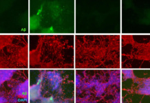
To get detailed images of how GLUT4 is transported and moves through the cell membrane, the researchers used high-resolution imaging to observe GLUT4 that had been tagged with a fluorescent dye.
The researchers then observed fat cells suspended in a neutral liquid and later soaked the cells in an insulin solution, to determine the activity of GLUT4 in the absence of insulin and in its presence.
In the neutral liquid, the researchers found that individual molecules of GLUT4 as well as GLUT4 clusters were distributed across the cell membrane in equal numbers. Inside the cell, GLUT4 was contained in balloon-like structures known as vesicles. The vesicles transported GLUT4 to the cell membrane and merged with the membrane, a process known as fusion.
After fusion, the individual molecules of GLUT4 are the first to enter the cell membrane, moving at a continuous but relatively infrequent rate. The researchers termed this process fusion with release.
When exposed to insulin, however, the rate of total GLUT4 entry into the cell membrane peaked, quadrupling within three minutes. The researchers saw a dramatic rise in fusion with release — 60 times more often on cells exposed to insulin than on cells not exposed to insulin.
After exposure to insulin, a complex sequence occurred, with GLUT4 shifting from clusters to individual GLUT4 molecules. Based on the total amount of glucose the cells took in, the researchers deduced that glucose was taken into the cell by individual GLUT4 molecules as well as by clustered GLUT4. The researchers also noted that after four minutes, entry of GLUT4 into the cell membrane started to decrease, dropping to levels observed in the neutral liquid in 10 to 15 minutes.
“The magnitude of this change shows just how important the regulation of this process is for the survival of the cell and for the normal function of the whole body,” said Joshua Zimmerberg, Ph.D., M.D., the paper’s senior author and director of the Eunice Kennedy Shriver National Institute of Child Health and Human Development (NICHD) Program in Physical Biology.
The research team next plans to examine the activity of glucose transporters in human fat cells, Zimmerberg said. “Understanding how insulin prepares the cell for glucose uptake may lead to ideas for stimulating this activity when the cells become resistant to insulin.”
Stenkula and Samuel W. Cushman, Ph.D., of NIDDK worked with NICHD investigators Vladimir A. Lizunov, Ph.D. and Zimmerberg to complete the research.
Story Source:
The above story is reprinted (with editorial adaptations by ScienceDaily staff) from materials provided by NIH/National Institute of Diabetes and Digestive and Kidney Diseases.
Journal Reference:
1. Karin G. Stenkula, Vladimir A. Lizunov, Samuel W. Cushman, Joshua Zimmerberg. Insulin Controls the Spatial Distribution of GLUT4 on the Cell Surface through Regulation of Its Postfusion Dispersal. Cell Metabolism, 2010; 12 (3): 250-259 DOI: 10.1016/j.cmet.2010.08.005


