


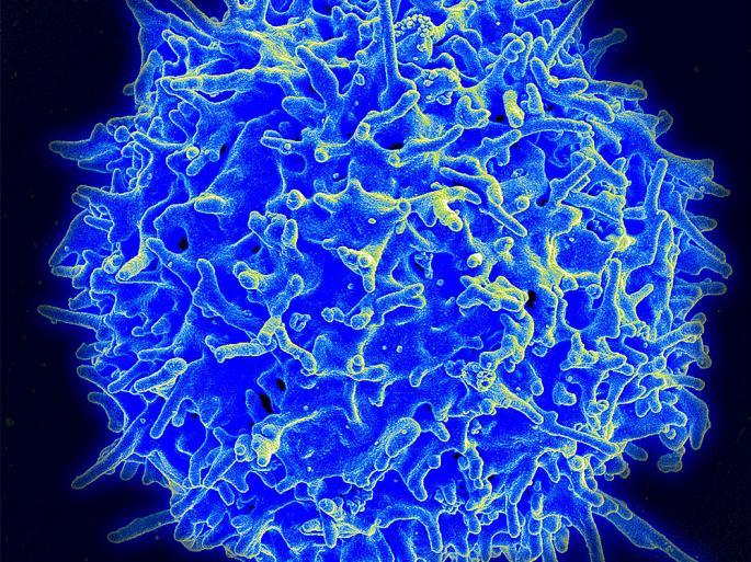
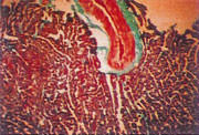
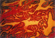
 The key to a healthy, functioning immune system rests with the thymus gland, a small organ lying just beneath the breastbone. The primary role of the thymus is to assist in the proliferation and differentiation of mature T-lymphocytes – cells that attack and kill viruses and bacteria. T-cells (Fig. 1) emerge from the bone marrow in an incomplete state. In order to function properly, immature T-cells migrate to the thymus gland where they are programmed into mature T-cells that orchestrate the immune response to attack and destroy invading viruses and cancer cells.
In our early twenties we have an abundance of well-trained, functioning T-cells that regulate the immune system and help the body fight off pathogens and disease.
After about age 20, the thymus begins to shrink (atrophy) as dying thymic cells are progressively replaced by fat and connective tissue.
By about age 40, output of thymic hormones has dropped significantly and T-cells have begun to lose their effectiveness. It is this gradual loss of functioning T-cells that is believed to be responsible for many of the age-related changes in the immune system that gradually rob the body of its ability to fight off infectious diseases, autoimmune disorders and cancer.
Antiaging Effects of Vital Cell in Rabbits
To evaluate the effects of Vital Cell on organ health, researchers conducted a two-year trial with two identical groups of rabbits. One group was treated with Vital Cell daily, and the second, untreated group served as a control. At the end of the study, the researchers compared the organs of both test groups of treated and untreated elderly rabbits to those of young, healthy juvenile rabbits.
When examining the treated rabbits the researchers noted that thymus glands of the old animals receiving Vital Cell retained the structure and functionality of glands normally seen only in young, health rabbits.
Conversely, the thymus glands of the old, untreated control rabbits were severely atrophied, weighing less than a third of their normal weight, and consisting primarily of inactive fat and connective tissues. Similar results were observed when the researchers compared tissues samples gathered from the brain, heart, liver, kidneys, spleen and other organs. In each case, the organs of Vital Cell-treated animals displayed the form and function of tissues normally only seen in younger subjects.
 |
 |
|
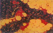
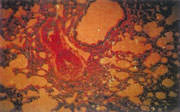
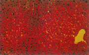
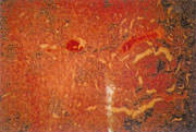
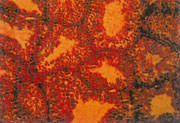
THYMUS – Aged rabbit
Weight is 1 gram, severe atrophy, heavy fatty infiltration |
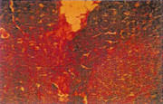
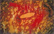
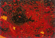
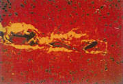
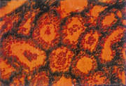
THYMUS – Treated rabbit
Weight is 2 grams, firm mass, slight atrophy, no fatty infiltrate |
Full Study with Color Slides
Observation of Effects of Vital Cell® Formula in Rabbits
Materials: 20 4-month-old, healthy purebred New Zealand rabbits, 10 males and 10 females each weighing 2kg. (Supplied by Shanghai Laboratory Animal Center of China Academy)
Grouping: Rabbits were divided randomly into two groups of 10 each, labeled Group A (the Vital Cell administration group) and Group B (the control group).
Feeding: Each rabbit in both groups was fed in separate cages in the same room. All were observed at room temperature. The feeding of rabbits is strictly defined according to time and quantity.
Observation Index and Methods
Method: After 2 years of treatment researchers randomly selected one male and one female rabbit from each group (A = treatment group, and B = control group). Researchers also selected one male and female rabbit from a group of 6-month-old, healthy purebred New Zealand rabbits weighing 2kg each. This group was labeled Group C (young, healthy controls).
Researchers randomly numbered all 6 rabbits and, after euthanizing, obtained tissue samples for examination with optical and electron microscopes. Tissues were prepared for optical microscopy by fixing with formalin, paraffin section, HE staining, weight elastic fiber staining of myocardium and coronary arteries, and NAP staining fixed by acetone for womb tissues.
Tissue samples for the electron microscope were fixed with 2.5% sporicidin, embedded with expoxide resin, prepped into ultra-thin sections and stained with acetate uranium and citrialuminum.
Observation Index: Researchers examined tissue samples from the thymus glands, hearts, lungs, livers, spleens, kidneys, brains, wombs and testes/womb of the 6 rabbits from Groups A, B, and C under optical microscope.
Researchers examined tissue samples from the livers of all 6 rabbits and testes of the 3 male rabbits from Group A, B, and C under electron microscope.
Results of Observation with Optical Microscope
Thymus gland: The thymus glands of the young, healthy rabbits in Group C was 3 grams, with all tissues and structures appearing normal. The weight of the thymus glands of the Vital Cell treated rabbits in Group A showed slight signs of atrophy, at 2 grams, retaining structure and functionality of glands observed in the younger rabbits. By contrast, the thymus glands of the old, untreated control rabbits in Group B were severely atrophied, weighing less than a third of their normal weight (1 gram) and consisting primarily of inactive fat and connective tissues.
|
|
|
|
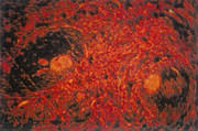
Thymus – Old rabbit
Thymus with severe atrophy, heavy fatty infiltration |
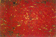
Thymus – Treated rabbit
Firm mass, only slight signs of atrophy |
Heart: Heart tissues revealed few alterations of the myocardium in the rabbits from Group A. Pathological changes in the coronary arteries were not obvious, and tunica intima was clear. The myocardium of the untreated older rabbits in Group B was fiberized, with extensive lipofuscin deposits and overgrown with collagen fibers and thickened and irregular intima. The myocardium and coronary arteries of the rabbits in Group C were normal.
 |
 |
|
Heart – Old rabbit
Sclerosis of coronary artery, thickened intima, fibrosis |
Heart – Treated rabbit
Slight myocardial fibrosis and unclear coronary sclerosis |
Lung: There were no obvious pathological changes in the blood vessels of the lungs of the rabbits in Group A. The walls of the arteriole of the lungs of the rabbits in Group B were thickened and the arterioles narrowed. The blood vessels of the lungs of the rabbits in Group C were normal.
 |
 |
|
Lung – Old rabbit
Thickening of small arteries, narrowing of arterioles |
Lung – Treated rabbit
No thickening or damage to small artery walls |
Liver: There was a little blood stagnation of the liver in the rabbits of Group A, but atrophy of hepatic cells was not obvious. Very little lipofuscin was found. Liver tissues from Group B revealed obvious signs of blood stagnation, atrophy of hepatic cells and significant lipofuscin deposits. The livers of the rabbits in Group C were normal.
 |
 |
|
Liver – Old rabbit
Stagnant blood, lipofuscin deposits in cells |
Liver – Treated rabbit
Absence of blood stagnation, no lipofuscin in cells |
Spleen: No signs of spleen blood stagnation in Group A, though walls of the central arteries of the splenic corpuscles were slightly thickened. There was blood stagnation of the spleen in Group B, and the walls of the central arteries of the splenic corpuscles thickened thus causing the arteries to become narrower. The rabbits in Group C were normal.
 |
 |
|
Spleen – Old rabbit
Vessel wall thickening of central arteries in splenic corpuscle |
Spleen – Treated rabbit
No vessel wall thickening of arteries in splenic corpuscle |
Kidney: In the rabbits of Group C, about 330 glomeruli were found under optical observation. In the treated rabbits in Group A there was no obvious change in structure and function of the kidneys, and 300 glomeruli were observed. In the older, untreated rabbits from Group B researchers observed significant changes of kidney structure, with thicker, narrowed arterioles and only 230 glomeruli.
 |
 |
|
Kidney – Old rabbit
Decrease in nephrons with few glomeruli |
Kidney – Treated rabbit
Slight decrease in nephrons |
Brain: Brain tissues from the young rabbits in Group C were normal. The meninx (brain covering) of the rabbits in Group A were normal, with slight signs of blood vessel expansion. In the older, untreated rabbits of Group B the meninx was thickened, and blood vessels of the lower arachnid membrane were expanded, showing evidence of blood stagnation and calcification.
 |
 |
|
Brain – Old rabbit
Calcification of arterial vessels, thickening of soft meninx |
Brain – Treated rabbit
No thickening of soft meninx, slight expansion of vessels |
Uterus: There was the enlargement of uterus in the rabbits of Group A, and the intima thickened. AKP staining showed (-) for the rabbits of Group A. There was atrophy of the womb in the rabbits of Group B. The womb became narrower, and the intima was thinner. AKP staining showed (+).
 |
 |
|
Uterus – Old rabbit
Thinning oftunica intima of the uterine lining |
Uterus – Treated rabbit
No thinning of tunica intima |
Testis: There was no obvious atrophy of the spermaduct in the rabbits of Group A, and there was a large amount of sperm. There was atrophy of the spermaducts in the rabbits of Group B and the sperm cells of various kinds were degenerated, reducing the number of sperm.
 |
 |
|
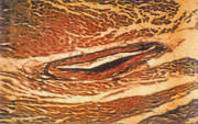
Testis – Old rabbit
Atrophy of the seminiferous tubules, reduced sperm |
Testis – Treated rabbit
No atrophy of the seminiferous tubules, healthy sperm |
Results of Observations with Electron Microscope
Liver: In Group A, the nuclei were either circular or oval, while the nuclear membrane was clear. The nucleoli had a thread-like structure. The nuclear chromatin was well distributed. The shape and volume of the mitochondria in Group A were similar to those in the healthy young rabbits. They were thick with numerous endoplasmic reticulum. Reticulation enlargement was not apparent.
In Group B, nuclei had an irregular shape, were separated and had lost their thread-like structure. Around the nucleoli, the chromatin had increased. Cytoplasmic mitochondria were swollen and shortened, and their shape was irregular. Dilatation of endoplasmic reticulum decreased. Lipofuscin deposits could be observed.
In Group C, the mitochondria were slightly enlarged, and other structures were normal.
Testis: Under electron microscope Group A showed no signs of atrophy of convoluted tubule, and there were no apparent degenerative changes in spermatoblasts. The nuclei and cell organs had a clear structure.
In Group B, the convoluted tubule was found to have shrunk under electron microscope. Spermatoblasts of different levels and sterol cells showed degenerative changes. Nuclear chromatin was condensed. Denaturation of intracytoplasmic cell organs and their obscure structure could be observed.
Results
In the older animals of Group B, the thymus glands, womb and testis displayed noticeable shrinkage. Coronary sclerosis, myocardial focal fibrosis, lipofuscin deposits and nephronal decrease were common. The use of an electron microscope revealed significant degenerative changes in the nucleoli and mitochondria of hepatic cells and convoluted tubule cells, accompanied by lipofuscin deposits.
In the Vital Cell treated rabbits in Group A, fewer pathological changes were apparent. In comparison to the young, healthy animals of Group C, the organic functions in Group A were normal.


Found it extremely disturbing that rabbits were murdered to test this product would therefore never use it.
I understand, not happy about it myself.
My cat just ate a rabbit, but it didn’t yield anything as useful as this study did. Thanks for the important data.
I appreciate the comprehensive analysis.