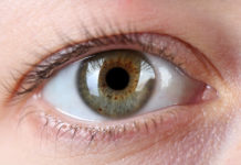
Researchers have developed a new bioengineered implant made of collagen protein from pig’s skin, which resembles the human cornea. In a pilot study, the implant restored vision to 20 people with diseased corneas, most of whom were blind prior to receiving the implant.
The promising results bring hope to those suffering from corneal blindness and low vision by providing an alternative to the transplantation of donated human corneas, which are scarce in countries where the need for them is greatest.
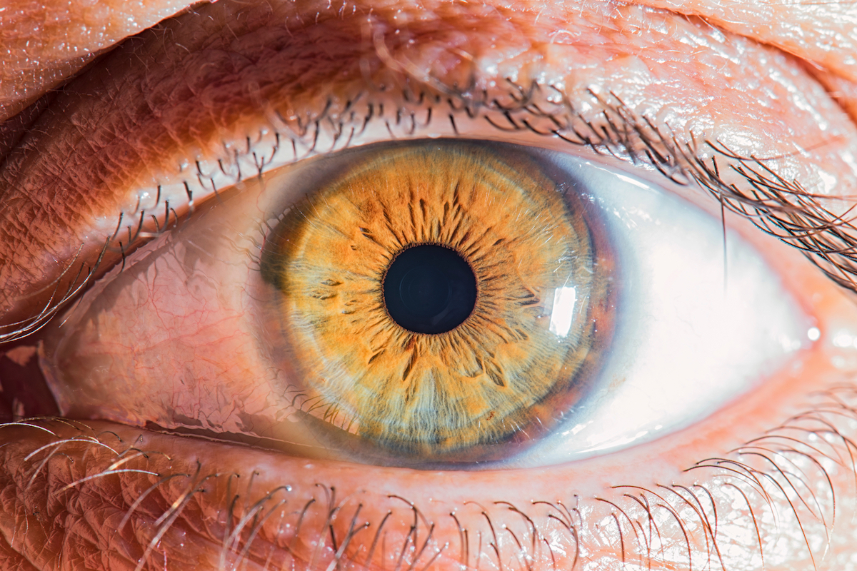
An estimated 12.7 million people around the world are blind due to their corneas, which is the outermost transparent layer of the eye, being damaged or diseased.
Their only way of regaining vision is to receive a transplanted cornea from a human donor. But just one in 70 patients receives a cornea transplant. Furthermore, most of those who need cornea transplants live in low and middle-income countries in which access to treatments is very limited.
“Safety and effectiveness of the bioengineered implants have been the core of our work, says Mehrdad Rafat, the researcher and entrepreneur behind the design and development of the implants.
“We’ve made significant efforts to ensure that our invention will be widely available and affordable by all and not just by the wealthy. That’s why this technology can be used in all parts of the world,” he says.
The cornea consists mainly of the protein collagen. To create an alternative to human cornea, the researchers used collagen molecules derived from pig skin that were highly purified and produced under strict conditions for human use. The pig skin used is a byproduct of the food industry, making it easy to access and economically advantageous. In the process of constructing the implant, the researchers stabilized the loose collagen molecules forming a robust and transparent material that could withstand handling and implantation in the eye.
While donated corneas must be used within two weeks, the bioengineered corneas can be stored for up to two years before use.
The researchers have also developed a new, minimally invasive method for treating the disease keratoconus, in which the cornea becomes so thin that it can lead to blindness. Today, a keratoconus patient’s cornea at advanced stage is surgically removed and replaced by a donated cornea, which is sewn into place using surgical sutures. This kind of surgery is invasive and only done at larger university hospitals.
“A less invasive method could be used in more hospitals, thereby helping more people. With our method, the surgeon doesn’t need to remove the patient’s own tissue. Instead, a small incision is made, through which the implant is inserted into the existing cornea,” says Neil Lagali, who has led the research group that has developed this surgical method.
No stitches are needed with this new surgical method. The incision in the cornea can be made with high precision thanks to an advanced laser, but also, when needed, by hand with simple surgical instruments.
The method was first tested on pigs and turned out to be simpler and potentially safer than a conventional cornea transplant.
The surgical method and the implants were used by surgeons in Iran and India, two countries where many people suffer from corneal blindness and low vision, but where there is a significant lack of donated corneas and treatment options. Twenty people who were either blind or on the verge of losing sight due to advanced keratoconus participated in the pilot clinical study and received the biomaterial implant.
The operations were free from complications; the tissue healed fast; and an eight-week treatment with immunosuppressive eye drops was enough to prevent rejection of the implant. With conventional cornea transplants, medicine must be taken for several years. The patients were followed for two years, and no complications were noted during that time.
The primary purpose of the pilot clinical study was to investigate whether the implant was safe to use. However, the researchers were surprised by what happened with the implant. The cornea’s thickness and curvature were restored to normal. At the group level, the participants’ sight improved as much as it would have after a cornea transplant with donated tissue.
Before the operation, 14 of the 20 participants were blind. After two years, none of them was blind any more.
Three of the Indian participants who had been blind prior to the study had perfect (20/20) vision after the operation.
A larger clinical study followed by market approval by regulatory authorities is needed before the implant can be used in healthcare. The researchers also want to study whether the technology can be used to treat more eye diseases, and whether the implant can be adapted to the individual for even greater efficacy.
Source: Mehrdad Rafat, Mahmoud Jabbarvand, Namrata Sharma, Maria Xeroudaki, Shideh Tabe, et al. Bioengineered corneal tissue for minimally invasive vision restoration in advanced keratoconus in two clinical cohorts. Nature Biotechnology, 2022; DOI: 10.1038/s41587-022-01408-w


