


 Swiss doctors have used cartilage cells harvested from patients’ own noses to successfully produce cartilage transplants for the treatment of the knees of 10 adults (aged 18-55 years) whose cartilage was damaged by injury. Two years after reconstruction, most recipients reported improvements in pain, knee function, and quality of life, as well as developing repair tissue that is similar in composition to native cartilage.
Every year, around 2 million people across Europe and the USA are diagnosed with damage to articular cartilage because of injuries or accidents.
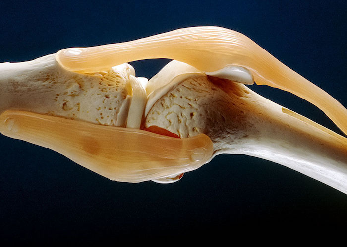
Articular cartilage is the tissue on the end of a bone that cushions the surface of the joint and is vital for painless movement. Because the tissue doesn’t have its own blood supply, it has limited capacity to repair itself once damaged, leading to degenerative joint conditions like osteoarthritis. Traditional methods to prevent or delay onset of cartilage degeneration following traumatic events like microfracture surgery don’t create the healthy cartilage needed to endure the forces of everyday movement. Even novel medical advances using patients’ own articular cartilage cells (chondrocytes) have been unable to predictably restore cartilage structure and function in the long term. As the population ages and people live longer, there is an urgent and growing need to develop an effective therapy to repair cartilage damage.
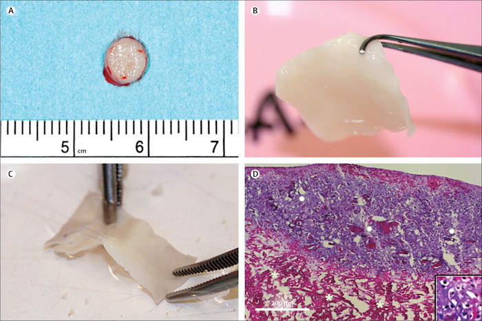
A team from the University Hospital Basel in Switzerland tried an alternative approach using engineered cartilage tissue grown from patients’ own cartilage cells from the nasal septum which have a unique capacity to grow and form new cartilage tissue. This phase 1 study included 10 patients with full-thickness cartilage lesions of the knee. The researchers extracted specimens from the nasal septum under local anesthetic using a minimally invasive procedure. The harvested cells were multiplied by exposing them to growth factors for 2 weeks. The expanded cells were then seeded onto collagen membranes and cultured for 2 additional weeks, generating a 30 x 40mm cartilage graft. The engineered graft was then cut into the right shape and used to replace damaged cartilage that was surgically removed from the recipient’s knee (Fig. 1).
MRI scans at 2 years revealed the development of new tissue with similar compositional properties of native cartilage. Moreover, nine recipients (one was excluded because of several independent sports injuries) reported substantial improvements in the use of their knee and in the amount of pain compared to before surgery. No adverse reactions were reported, although two serious adverse events unrelated to the procedure were recorded — an independent injury in the opposite knee and new cartilage lesions at other locations in the treated knee.
The researchers say that the small number of participants and the relatively short follow-up time will mean further studies will be needed. Similar to other early phase surgical studies, the trial did not involve a control group, so other studies will be needed to establish a comparison in effectiveness with currently available treatments, and to assess the possible bias of a placebo effect.
Source: Marcus Mumme, Andrea Barbero, Sylvie Miot, Anke Wixmerten, Sandra Feliciano, Francine Wolf, Adelaide M Asnaghi, Daniel Baumhoer, Oliver Bieri, Martin Kretzschmar, Geert Pagenstert, Martin Haug, Dirk J Schaefer, Ivan Martin, Marcel Jakob. Nasal chondrocyte-based engineered autologous cartilage tissue for repair of articular cartilage defects: an observational first-in-human trial. The Lancet, 2016; 388 (10055): 1985 DOI: 10.1016/S0140-6736(16)31658-0


